Magnification
-the object is the thing being viewed.
-the image is the appearance of the object when viewed under the microscope.
-magnification is how many times bigger the image is compared to the object.
-the image is the appearance of the object when viewed under the microscope.
-magnification is how many times bigger the image is compared to the object.
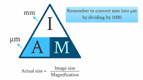
-remember, before calculating, make sure the image and the object have the same units.
Resolution
-the resolving power/resolution is the minimum distance apart that two objects can be in order for the to appear as separate items.
-greater resolution, means greater clarity.
-greater resolution, means greater clarity.
Cell Fractionation
-cell fractionation is the process of breaking up a cell into its individual organelles.
1) place the cells in a cold (to reduce enzyme activity that may break down organelles), isotonic (to prevent organelles from bursting or shriveling due to different water potentials causing osmosis) and buffered (to maintain a constant pH) solution.
2) cells are then put in a homogeniser or vibrated, in order to break the plasma membrane and release the organelles.
3) the mixture (called the homogenate) is then filtered through a gauze to separate away any larger debris or complete cells- organelles are small so can pass through the gauze.
4) the organelles are then separated by ultracentrifugation. The filtered homogenate is put in a tube to be spun in the ultracentifuge. The speeds it is spun creates a centrifugal force.
5) at lower speeds, the heaviest (biggest mass) organelles (e.g. nuclei) are flung to the bottom of the tube, forming a sediment called a pellet.
6) the supernatent (remaining fluid) is poured into another tube for the process to be done at a higher speed (this time mitochondria form a pellet).
7) this is repeated until all the organelles are separated, allowing you to study them individually.
Questions
1) An object is 100nm long, and when under a microscope, is 10mm long. What is the magnification? (2 marks)
2) What is the difference between magnification and resolution? (2 marks)
3) Describe the process of cell fractionation, explaining why each step is done. (8 marks)
2) What is the difference between magnification and resolution? (2 marks)
3) Describe the process of cell fractionation, explaining why each step is done. (8 marks)
